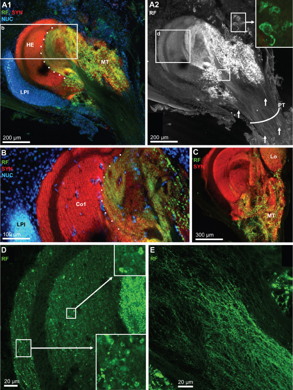
Figure 16.
Details of the hemielliposid body and the medulla terminalis; triple labeling for synapsin immunoreactivity (SYN; red), RFamide-like immunoreactivity (RF; green), plus the nuclear marker (NUC) shown in conventional fluorescence combined with the Apotome structured illumination technique (A1, B) and confocal laser scan microscopy (A2, C-E). A1: a massif cluster of lateral protocerebral interneurons (LPI) is associated with the hemiellipsoid body (HE). The dotted line demarks the field of strong innervation with peptidergic neurites. The medulla terminalis (MT) is filled with a loose meshwork of peptidergic fibers. The boxed area is shown at a higher magnification in B. A2: same specimen as in A1 but visualized with confocal microscopy and showing only the RFamide channel. Arrows identify single, peptidergic fibers within the protocerebral tract (PT). Peptidergic interneurons are associated with the medulla terminalis (inset). The boxed areas d and e are shown at a higher magnification in D and E. B: Already at moderate magnification, the core neuropil (Co1) displays a layered appearance. The dotted line demarks the field of strong innervation with peptidergic neurites. The cluster of lateral protocerebral interneurons is also visible (LPI) as are single cell nuclei (presumably endothelial cells of blood vessels) within the core neuropil. C: The medulla terminalis (MT) is filled with a loose meshwork of peptidergic fibers. D: Higher magnification of the boxed area in A2. Both the cap and core 1 neuropils are filled with numerous very small RFir profiles some of which are arranged in a string (see insets) suggesting an intense peptidergic innervation of these neuropils. E: higher magnification of the boxed area in A2 showing a meshwork of very fine peptidergic fibers within the olfactory globular tract close to its entrance into the medulla terminalis/hemiellipsoid body complex.

