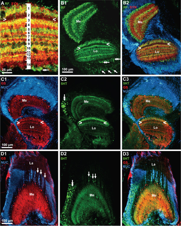
Figure 19.
A, B: lobula (see A) and medulla (Me) and lobula (Lo; B1, B2); triple labeling for synapsin immunoreactivity (SYN; red), RFamide-like immunoreactivity (RF; green), plus the nuclear marker (NUC) shown in conventional fluorescence combined with the Apotome structured illumination technique. In the medulla, RFir is localized in three distinct parallel layers. In the lobula (see A), fourteen different layers can be recognized with this technique. Arrowheads label a conspicuous weakly labelled layer. Double arrows identify proximally located, large RFir profiles associated with the lobula. Arrows identify RFir somata of visual interneuons. C1–C3: lobula (Lo) and medulla (Me); triple labeling for serotonin immunoreactivity (5HT; green), glutamine synthetase-like immunoreactivity (RF; red), plus the nuclear marker (NUC; blue) shown in conventional fluorescence combined with the Apotome structured illumination technique. Arrowheads label a conspicuous weakly labelled layer (compare B2). D1–D3: lamina (Lo) and medulla (Me); triple labeling for serotonin immunoreactivity (5HT; green), glutamine synthetase-like immunoreactivity (RF; red), plus the nuclear marker (NUC; blue) shown in conventional fluorescence combined with the Apotome structured illumination technique. Arrows in D1 label the course of fiber bundles that link the lamina and the medulla. Arrows in D2 identify serotonergic somata located between the lamina and the medulla. Double arrows in D2 label serotonergic neurons associated laterally with the medulla.

