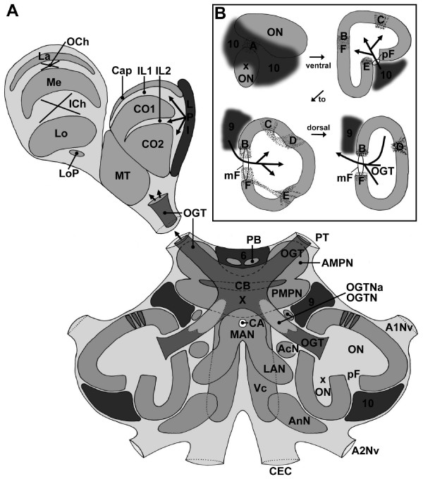
Figure 2.
A: Idealized schematic drawing of the C. clypeatus brain (dorsal view) compiled from ca. 4–5 successive sections (80 μm) of several animals at a mid horizontal level. B: Schematic cartoons of ventral to dorsal sections of the main olfactory lobe (ON) and the side olfactory lobe (xON) to show the localization of the median (mF) and posterior foramina (pF) and the patches of non-columnar neuropil (dotted areas labeled with letters A-F). Arrows labeled 9 and 10 show the input of olfactory interneurons. The arrow labeled OGT shows the exit of the olfactory globular tract. Abbreviations: 6, 9, 10 cell clusters 6, 9, 10, A1Nv nerve of antenna 1, A2Nv nerve of Antenna 2, AcN acessory lobe/neuropil, AMPN anterior medial protocerebral neuropil, AnN antenna 2 neuropil, CA cerebral artery, Cap cap neuropil of the hemiellipsoid body, CB central body, CEC circumesophageal connectives, CO1, CO2 core neuropils 1 and 2 of the hemiellipsoid body, ICh inner optic chiasm, IL1, IL2 intermediate layers 1 and 2 of the hemiellipsoid body, La Lamina (lamina ganglionaris), LAN lateral antenna 1 neuropil, Lo Lobula (medulla interna), LoP Lobula "plate", LPI lateral protocerebral interneurons, MAN median antenna 1 neuropil, Me Medulla (medulla externa), mF median foramen, MT Medulla terminalis, OCh outer optic chiasm, OGT olfactory globular tract, OGTN olfactory globular tract neuropil, OGTNa accessory olfactory globular tract neuropil, ON olfactory lobe/neuropil, PB protocerebral bridge, pF posterior foramen, PMPN posterior medial protocerebral neuropil, PT protocerebral tract, VC ventral neuropil column, X chiasm of the olfactory globular tract.

