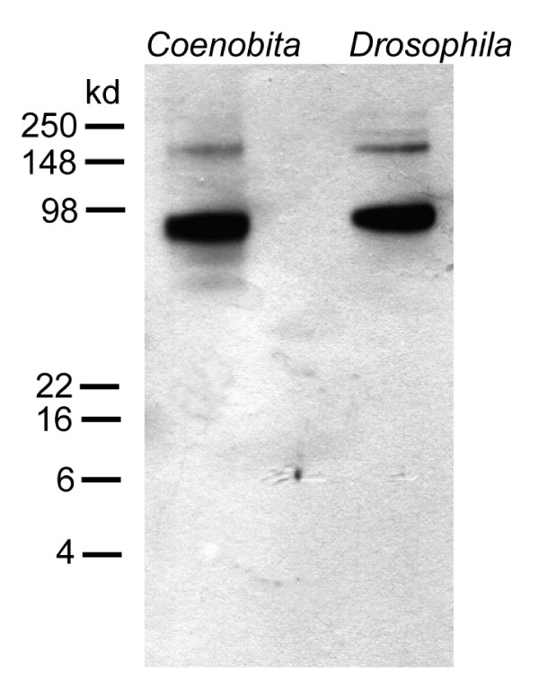
Figure 20.
Western Blot analysis of the SYNORF1 antibody (anti-synapsin) comparing brain tissue of Drosophila and Coenobita. The antibody provided identical results for both species staining one strong band around 80–90 kDa and a second weaker band slightly above 148 kDa. This pattern closely resembles the results obtained in the original publication in which the antibody was characterized for Drosophila (Klagges et al. 1996).

