
Figure 4.
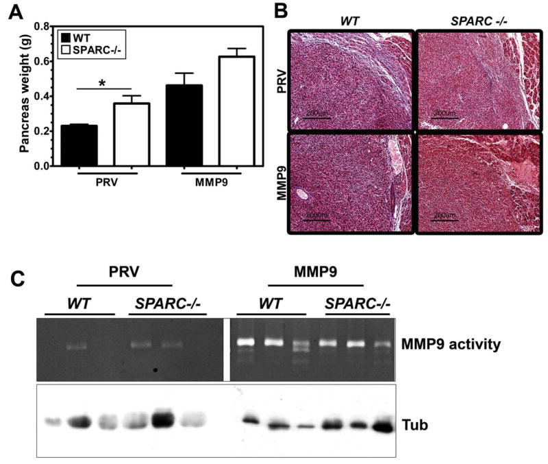
Forced expression of MMP9 or the absence of host SPARC increases PAN02 tumor growth. A) PAN02-PRV (PRV) and PAN02-MMP9 (MMP9) cells (5 × 10⁵) were implanted orthotopically in the pancreas of age- and sex-matched SPARC-/- and WT mice (n= 6/group). After 6 weeks, the entire pancreas including tumor was weighed (g). A comparison of mean +/- SD pancreas weight is shown (* p<0.05, Student's t test). One way ANOVA with Tukey's multiple comparison test revealed significant differences across tumor groups as well. PAN02-PRV tumors in WT mice were significantly smaller than PAN02-MMP9 tumors grown in WT mice (p<0.01) or SPARC-/- mice (p<0.001) while PAN02-PRV tumors in SPARC-/- mice were significantly different from PAN02-MMP9 in SPARC-/- mice (p<0.001). B) Masson's trichrome staining of paraffin-embedded tumors revealed decreased collagen fibers (blue) in tumors grown in SPARC-/- mice compared with those in WT mice. Forced expression of MMP9 resulted in an even further reduction in collagen deposition in response to tumor growth. C) Tumor extracts from PAN02-PRV (PRV) and PAN02-MMP9 (MMP9) grown in WT and SPARC -/- mice were analyzed by gelatin zymography. The bands corresponding to MMP9 are indicated. Western blot analysis of tubulin in equivalent amount of extracts was performed as a loading control.