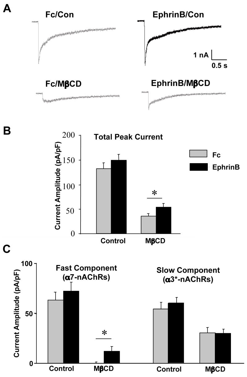
Figure 4.
Effects of ephrinB1 treatment on nicotine-induced whole-cell currents. (A) Representative traces of nicotine-induced whole-cell responses in E14 neurons after treatment with Fc/Ab as a control (Fc, left) or with ephrinB1-Fc/Ab (EphrinB, right) and then incubated in culture medium (Con, top) or extracted with MβCD to extract cholesterol and disperse lipid rafts (MβCD, bottom). (B) Quantification of the peak currents, normalized for capacitance to correct for differences in cell size. (C) Quantification of the peak current components contributed by α7-nAChRs (left) and α3*-nAChRs (right), separated based on decay rates (fast vs. slow, respectively). Values represent the mean ± SEM of 12–18 cells. Asterisks, p < 0.05. The ephrinB1 treatment does not significantly enhance the nicotine-induced whole-current but does partially protect the rapidly decaying α7-nAChR component of the response against MβCD-induced loss.

