
Figure 1. Induction of DNA laddering following infection with BHV-1 or UV light treatment.
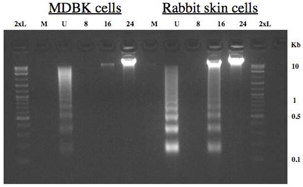
RS or MDBK cells were mock infected (M), UV treated (U), or infected with wt BHV-1 (MOI = 1) for 8, 16, or 24 hours. UV treated cells were exposed to UV light from a trans-illuminator for 30 sec (312 nm), fresh media was added to each well, and cultures were incubated for 4 h at 37 °C. After virus inoculation, cells were incubated for 1 h at 37°C, monolayers were rinsed twice with CMF-PBS, and EMEM supplemented with 5% FCS was added to each well. DNA was electrophoresed through a 2% agarose gel in 1x TAE. A 2-log molecular weight DNA marker (New England Biolabs) (2xL) was used as a DNA marker.