
Figure 2. Induction of DNA laddering following infection with the bICP0 null mutant.
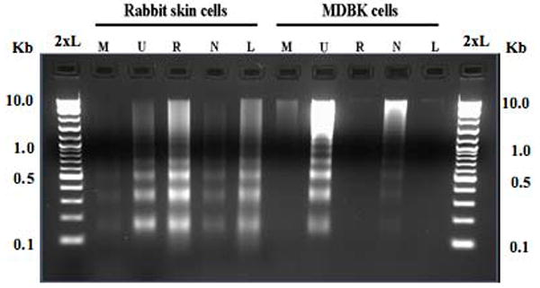
RS or MDBK cells were mock infected (M), UV treated (U), or infected with the bICP0-rescued virus (R), bICP0 null mutant (N), or the LR mutant (L). A MOI = 1 was used for all viruses. After virus inoculation, cells were incubated for 1 h at 37°C, monolayers were rinsed twice with CMF-PBS, and EMEM supplemented with 5% FCS was added to each well. Two days after plating, media was removed from mock-infected RS or MDBK cells, and these cells were exposed to UV light as described in Figure 1. At 48 h after infection, cells were collected and small molecular weight DNA prepared as described in materials and methods. DNA was electrophoresed along with a 2-log molecular weight DNA marked (New England Biolabs) through a 2% agarose gel in 1x TAE (2xL).