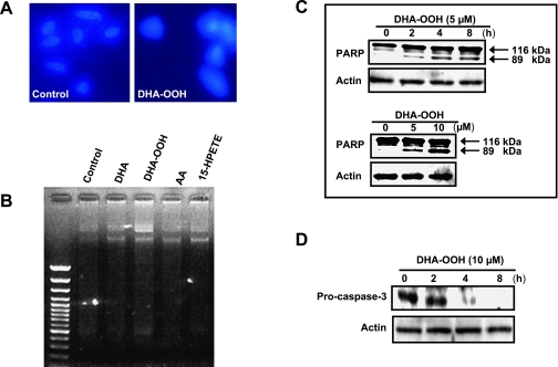
Fig. 2.
Induction of apoptotic cell death. A, Nuclei condensation in SH-SY5Y cells exposed to 10 µM DHA-OOH. SH-SY5Y cells were stained with Hoechst 33258, and examined by fluorescence microscopy. Left panel, control. Right panel, DHA-OOH treatment. B, DNA fragmentation in SH-SY5Y cells exposed to 10 µM DHA-OOH for 4 h. Nucleosomal DNA fragmentation was visualized by agarose gel electrophoresis. C, PARP cleavage in SH-SY5Y cells exposed to DHA-OOH. The cleavage of PARP was analyzed by Western blotting. Upper panel, time-dependent cleavage of PARP. Lower panel, dose-dependent cleavage. D, Analysis of caspase-3 expression. The cells were treated with 10 µM DHA-OOH for 0–8 h. Expression of pro-caspase 3 was detected by western blotting using caspase-3 antibody (not recognizing cleaved form of caspase-3 in the western blot).

