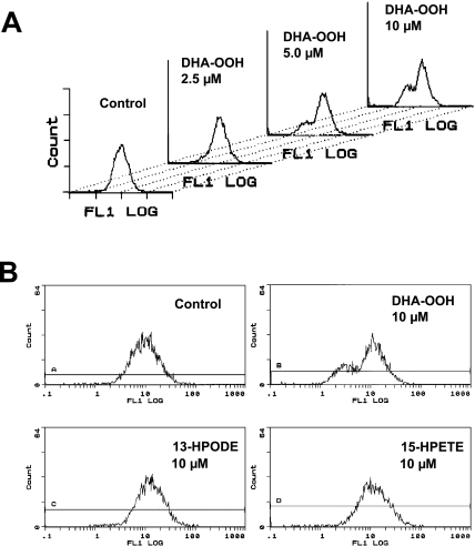
Fig. 4.
Loss of mitochondrial membrane potential. A, SH-SY5Y cells were treated with different concentration of DHA-OOH (0, 2.5, 5, 10 µM). DiOC6 was added to the culture medium for a further 30 min. The fluorescence intensity of DiOC6 in cells was analyzed by flow cytometry. B, As described in A, the effect of 13-HPODE and 15-HPETE to mitochondria membrane potential were also measured compared to DHA-OOH.

