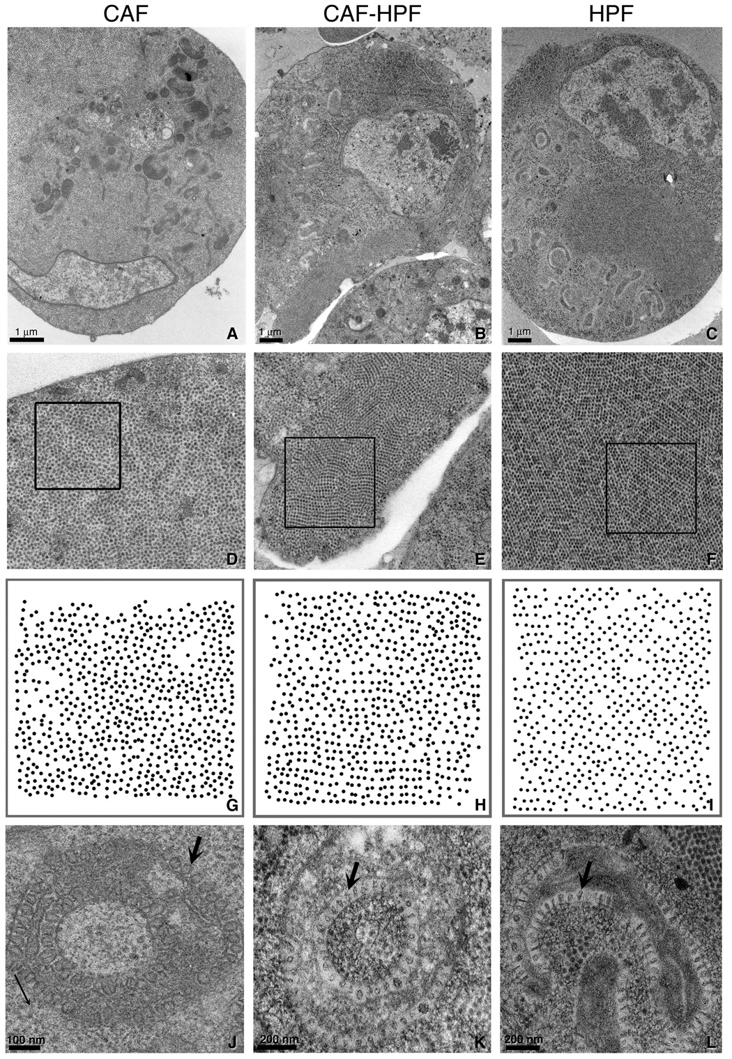
Figure 1. Comparison of preparation methods for preserving ultrastructure in FHV infected DL1 cells.

Late stage FHV infected DL1 cells prepared by conventional aldehyde fixation and embedding methods (CAF) (A, D, G, J), fixed and HPF (CAF-HPF) (B, E, H, K) and HPF alone (C, F, I, L) were examined by thin section EM. A higher magnification view of the aggregates of FHV in each DL1 cell in A (3x), B (3.5x) and C (3.5x) are shown in D, E and F respectively. The diameter of an individual FHV is 30 nm. A plot of the positions of the viruses centers in the boxed areas in D, E, and F are displayed in G, H and I, respectively and illustrating a more crystalline arrangement in H and I as compared with G. (J–L) Morphology of an appropriated mitochondrion in each preparation method. An arrow in each micrograph points to a spherule. Note how similar the mitochondrial morphologies are in the insets in K and L as compared to that in J, which appears empty and swollen.
