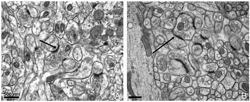
Figure 2. Comparison of conventionally aldehyde fixed versus conventionally aldehyde fixed and high pressure frozen brain tissue.

(A) Conventionally prepared cerebellar tissue. (B) Cerebellum slices that had been chemically fixed prior to HPF. The black arrows point to a postsynaptic density (PSD) that appears thicker in (B) than in (A).
