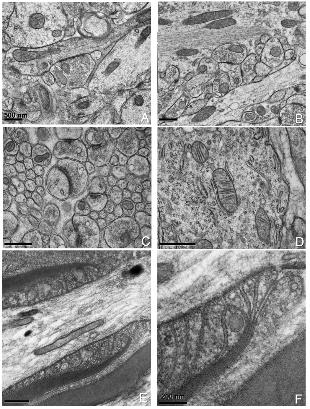
Figure 3. Examples of nervous tissue prepared by a combined chemical fixation and HPF.

(A, B) Hippocampal slices that have been chemically fixed prior to HPF. (C) Cerebellar slices showing good preservation of synaptic vesicles and PSD and (D) mitochondria with attached smooth and rough endoplasmic reticulum. Note the ribosomes in the background of (D). (E) Spinal root prepared by combined chemical fixation and HPF. Note the smoothness of the membranes and the level of detail in the paranodal loops and axonal-glial junctions. A 3x magnification is shown in (F).
