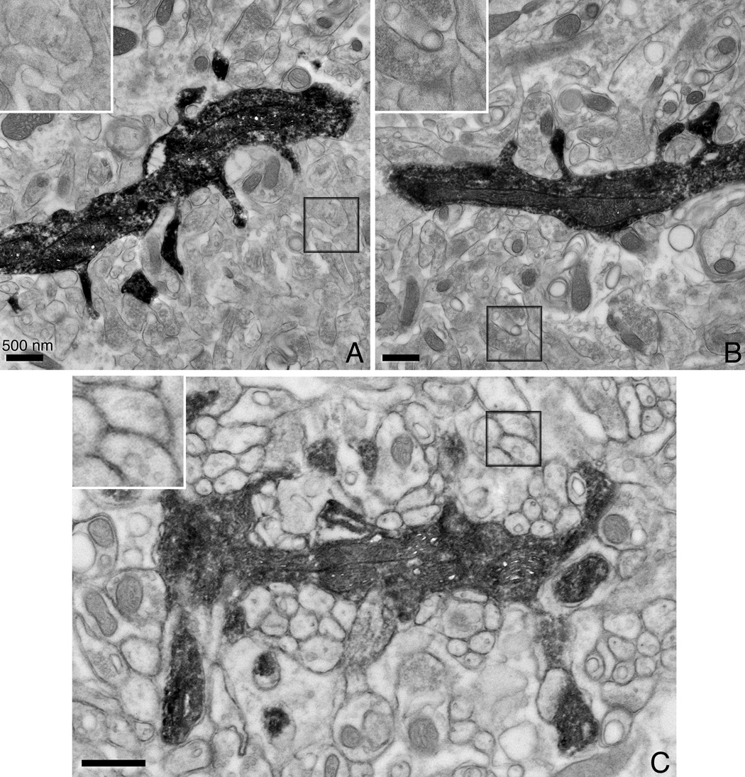
Figure 5. Thin section EM images of chemically fixed neurons that have been filled with Lucifer Yellow, photooxidized and HPF.

Conventionally prepared (A) and aldehyde fixed/HPF (B) hippocampal neurons. The Lucifer Yellow-DAB-osmium precipitate provides selective staining of the filled neuron with its dendrites. Note that while the ultrastructure of (A) and (B) are comparable, the delineation and preservation of the cell membranes is better in (B). (C) A photoconverted cerebellar Purkinje neuron also shows good ultrastructural preservation with chemical fixation/HPF. All scale bars correspond to 500 nm. The inset shown in each image is the boxed area shown at twice the magnification.
