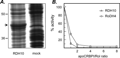
FIGURE 1.
Expression of RDH10 in Sf9 cells and effect of apoCRBPI on RDH10 retinol dehydrogenase activity. A, Sf9 cells were infected with recombinant (RDH10-His6) or wild-type (mock) Baculovirus vector and microsomes were isolated by differential centrifugation 3 days later. Microsomal proteins were separated by SDS-PAGE and the proteins were visualized by staining with Coomassie R-250. The position of RDH10 protein is indicated by an arrow. B, the reactions were carried out with microsomal preparation of RDH10 expressed in Sf9 cells (5 μg) in the presence of 1 mm NAD+ for 15 min at 37 °C. RoDH4 expressed in microsomes of Sf9 cells (5 μg) was used for comparison. The concentration of all-trans-retinol (Rol) was 1μm, whereas the concentration of apoCRBPI was varied from 0 to 8 μm. The results shown are mean ± S.D. of three independent experiments and were reproduced with three different preparations of apoCRBPI.

