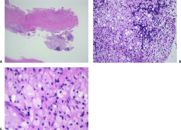
Figure 2.
(A) Lower magnification (4×) photomicrograph of the cyst wall and its contents: cuboidal epithelium (two-layered in most areas) with evidence of keratinization and considerable sclerosis. (B) (20×) Photomicrograph of the cyst wall: there are cholesterol clefting and foamy macrophages associated with the cyst. (C) (60×) Photomicrograph with high magnification: note the glandular structures containing proteinaceous material.

