
Fig. 1. Yolk sac-derived blood cells from Brg1fl/fl:Tie2-Cre+/0 embryos are scarce and morphologically abnormal by E9.5.
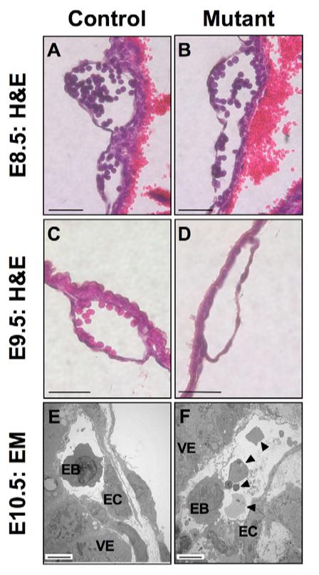
(A,B) Histological sections of Brg1fl/fl (A) and Brg1fl/fl:Tie2-Cre+/0 (B) yolk sac vessels filled with hematopoietic blood cell progenitors, from littermate E8.5 embryos. (C,D) Histological sections of a Brg1fl/fl yolk sac vessel (C) and a Brg1fl/fl:Tie2-Cre+/0 yolk sac vessel (D). The mutant vessel is devoid of embryonic blood cells. Scale bars in A-D: 40 μm. (E,F) Transmission electron micrographs of E10.5 Brg1fl/+ (E) and Brg1fl/fl:Tie2-Cre+/0 (F) yolk sac blood vessels. Arrowheads indicate abnormal embryonic blood cells and blood cell fragments. EB, embryonic blood cell; EC, endothelial cell; VE, visceral endoderm. Scale bars in E,F: 5 μm.