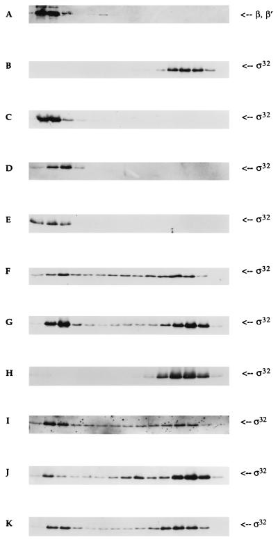
Figure 2.
Western blot analysis of glycerol gradient sedimentation of sigma factors and core RNAP. The concentration of each protein in all experiments was 100 nM. The sedimentation pattern of both σ32 and core RNAP was determined using polyclonal antibodies [gift of C. Gross (University of California, San Francisco) and Y. N. Zhou (National Institutes of Health, Bethesda, MD)]. The left-most region of each panel represents the bottom of the tube (35% glycerol). A typical sedimentation pattern of β and β′ subunits of core RNAP with σ32 (A); σ32 only (B); σ32 with core RNAP (C); E81G with core RNAP (D); P74R with core RNAP (E); Q80R with core RNAP (F); σ32 + σ70 with core RNAP (G); Q80R + σ70 with core RNAP (H); P74R + σ70 with core RNAP (I); E81G + σ70 with core RNAP (J); and Q80N with core RNAP (K).

