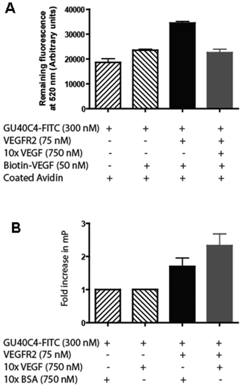
Figure 5.

Formation of a peptoid•VEGFR2 ECD•VEGF complex. (A) The reagents indicated in the figure were incubated in the wells of biotin-VEGF captured avidin-coated plates. After washing, the intensity of the retained fluorescence at 520 nm was monitored to score retention of GU40C4-FITC. A significant increase in GU40C4-FITC retention above background (first bar) was observed only in the presence of VEGFR2 ECD (third bar). This retention was abolished by excess soluble VEGF. (B) Fluorescence anisotropy experiment monitoring the change in anisotropy evinced by GU40C4-FITC in the presence of the indicated reagents.
