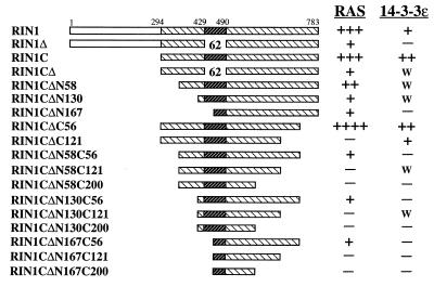
Figure 3.
Interaction of RIN1 with H-RAS and 14-3-3ɛ. Labeled RIN1 was analyzed separately for normalization. Bound protein was analyzed by SDS/PAGE and autoradiography. Binding above background (GST or MBP alone) was assessed. Multiple +’s indicate strong binding; “w” indicates weak binding (<2-fold above background).

