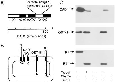
Figure 4.
Membrane topology of DAD1. (A) The predicted membrane spans of DAD1 are shown as solid bars. The location of the peptide (AsnProGlnAsnLysAlaAspPheGlnGlyIleSerProGluArg) used for antibody production is shown using the one letter code for amino acids. Potential trypsin sites (Lys and Arg) in DAD1 are indicated by asterisks, and potential chymotrypsin sites (Tyr, Phe, and Trp) are indicated by diamonds. (B) A model for the membrane topology of DAD1 locates both the N terminus and the antigenic peptide on the cytoplasmic face of the membrane. OST48 (18) and ribophorin I (R I) (19, 20) are integral membrane proteins with lumenal N termini (N). (C) Aliquots of PK-RM were incubated at 37°C for 3.75 h with either trypsin (60 μg/ml) or chymotrypsin (Chymo.) (60 μg/ml) with or without Triton X-100. Each sample was divided for subsequent SDS/PAGE using a 15% polyacrylamide gel for the DAD1 immunoblot or 9% polyacrylamide gels for the OST48 and ribophorin I (R I) immunoblots. The blots were probed with antibodies specific for the lumenal domains of ribophorin I and OST48. Proteolysis of ribophorin I yields a 50-kDa fragment (R I*) in the absence of detergent.

