Abstract
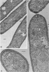
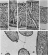
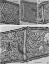
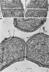
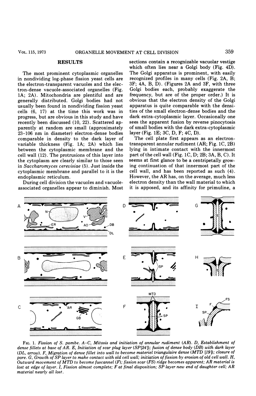
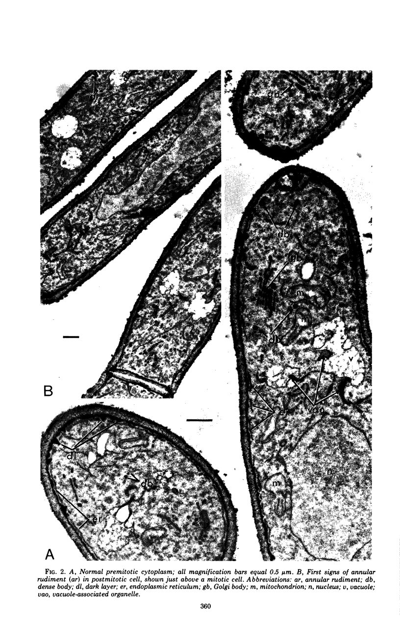
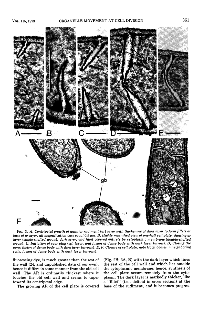
Electron microscopy of dividing fission yeast cells shows establishment of an annular rudiment (AR) of electron-transparent material under the old cell wall as the first sign of elaboration of the cell plate. The AR grows centripetally, finally closing at the mid-point of the cell. During the inward growth of the AR it is thickened by addition of denser material which becomes the scar plug after fission; the electron-transparent material is lost at fission. Lying always between the cytoplasmic membrane and the cell wall is a dark layer of variable thickness. This layer becomes markedly thickened into a fillet at the base of the centripetally growing cell plate. The fission process begins after the cell plate is completely elaborated. One striking feature of fission is the migration of dense material from the fillet at the base of the cell plate outwardly through the matrix of the cell wall to its final resting place as a dark ring, a “fuscannel,” adjacent to the fission scar. The inclusion of Golgi bodies in many sections suggests their involvement in cell plate elaboration, presumably through production of the dense bodies which are seen to fuse with the dark layer proximal to the growing cell plate.
Full text
PDF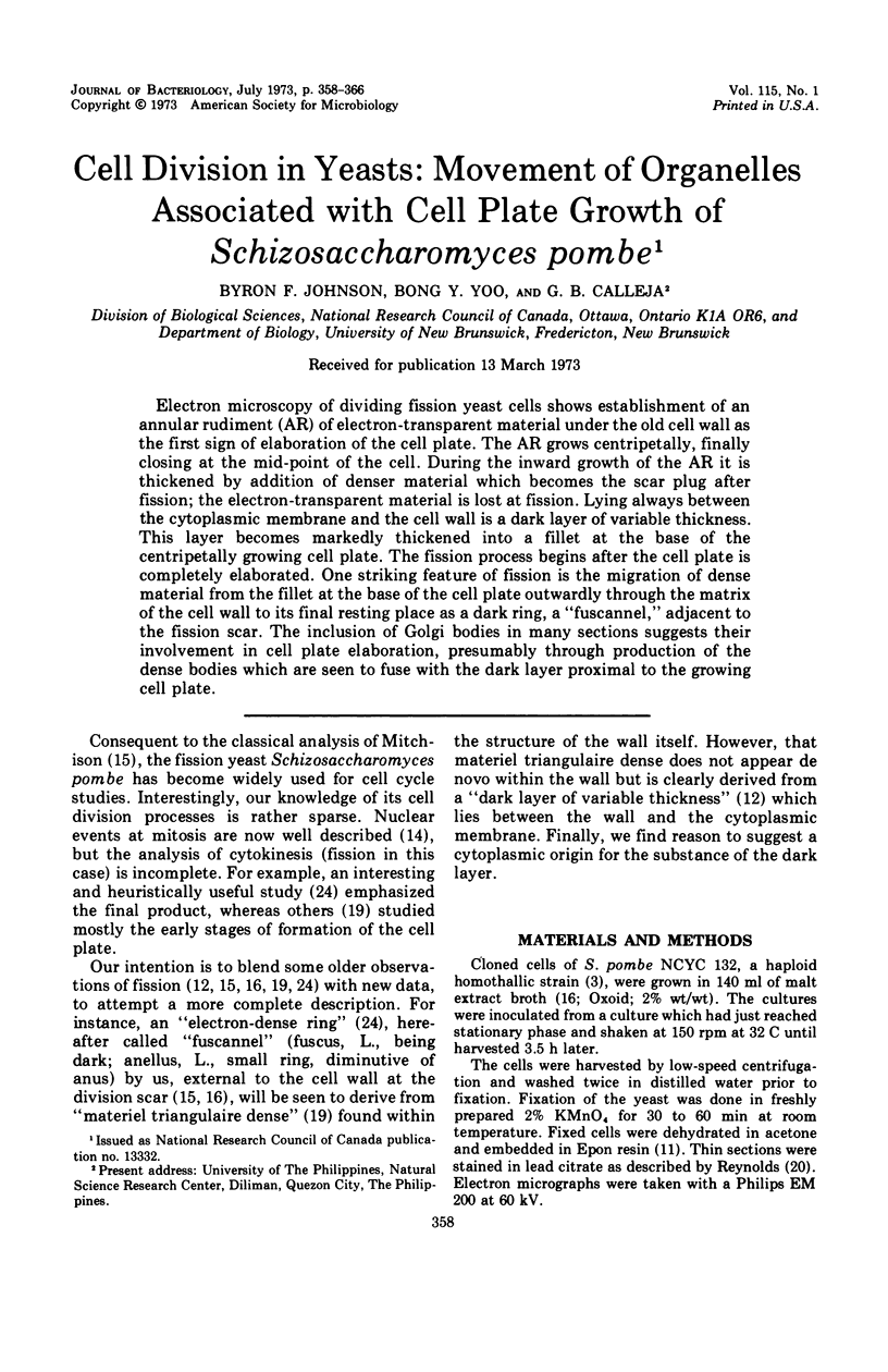
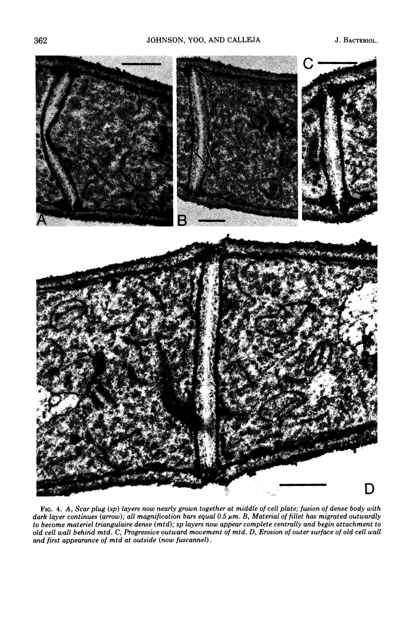
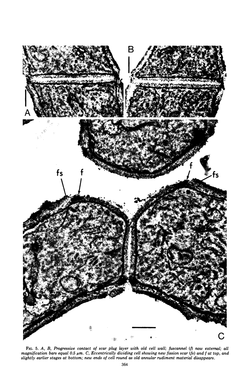
Images in this article
Selected References
These references are in PubMed. This may not be the complete list of references from this article.
- BARTON A. A. Some aspects of cell division in saccharomyces cerevisiae. J Gen Microbiol. 1950 Jan;4(1):84–86. doi: 10.1099/00221287-4-1-84. [DOI] [PubMed] [Google Scholar]
- CONTI S. F., NAYLOR H. B. Electron microscopy of ultrathin sections of Schizosaccharomyces octosporus. I. Cell division. J Bacteriol. 1959 Dec;78:868–877. doi: 10.1128/jb.78.6.868-877.1959. [DOI] [PMC free article] [PubMed] [Google Scholar]
- Calleja G. B., Johnson B. F. Flocculation in a fission yeast: an initial step in the conjugation process. Can J Microbiol. 1971 Sep;17(9):1175–1177. doi: 10.1139/m71-187. [DOI] [PubMed] [Google Scholar]
- Ghosh B. K. Grooves in the plasmalemma of Saccharomyces cerevisiae seen in glancing sections of double aldehyde-fixed cells. J Cell Biol. 1971 Jan;48(1):192–197. doi: 10.1083/jcb.48.1.192. [DOI] [PMC free article] [PubMed] [Google Scholar]
- Johnson B. F. Autoradiographic analysis of regional cell wall growth of yeasts. Schizosaccharomyces pombe. Exp Cell Res. 1965 Sep;39(2):613–624. doi: 10.1016/0014-4827(65)90064-9. [DOI] [PubMed] [Google Scholar]
- Johnson B. F. Morphometric analysis of yeast cells. Adult cell volume of Saccharomyces cerevisiae. Exp Cell Res. 1965 Sep;39(2):577–583. doi: 10.1016/0014-4827(65)90059-5. [DOI] [PubMed] [Google Scholar]
- Johnson B. F. Morphometric analysis of yeast cells. II. Cell size of Schizosaccharomyces pombe during the growth cycle. Exp Cell Res. 1968 Jan;49(1):59–68. doi: 10.1016/0014-4827(68)90519-3. [DOI] [PubMed] [Google Scholar]
- Kopecka M. Dictyosomes in the yeast Schizosaccharomyces pombe. Antonie Van Leeuwenhoek. 1972;38(1):27–31. doi: 10.1007/BF02328074. [DOI] [PubMed] [Google Scholar]
- LUFT J. H. Improvements in epoxy resin embedding methods. J Biophys Biochem Cytol. 1961 Feb;9:409–414. doi: 10.1083/jcb.9.2.409. [DOI] [PMC free article] [PubMed] [Google Scholar]
- MACLEAN N. ELECTRON MICROSCOPY OF A FISSION YEAST, SCHIZOSACCHAROMYCES POMBE. J Bacteriol. 1964 Nov;88:1459–1466. doi: 10.1128/jb.88.5.1459-1466.1964. [DOI] [PMC free article] [PubMed] [Google Scholar]
- MITCHISON J. M. The growth of single cells. I. Schizosaccharomyces pombe. Exp Cell Res. 1957 Oct;13(2):244–262. doi: 10.1016/0014-4827(57)90005-8. [DOI] [PubMed] [Google Scholar]
- Matile P., Wiemken A. The vacuole as the lysosome of the yeast cell. Arch Mikrobiol. 1967 Feb 20;56(2):148–155. doi: 10.1007/BF00408765. [DOI] [PubMed] [Google Scholar]
- McCully E. K., Robinow C. F. Mitosis in the fission yeast Schizosaccharomyces pombe: a comparative study with light and electron microscopy. J Cell Sci. 1971 Sep;9(2):475–507. doi: 10.1242/jcs.9.2.475. [DOI] [PubMed] [Google Scholar]
- Osumi M., Sando N. Division of yeast mitochondria in synchronous culture. J Electron Microsc (Tokyo) 1969;18(1):47–56. [PubMed] [Google Scholar]
- REYNOLDS E. S. The use of lead citrate at high pH as an electron-opaque stain in electron microscopy. J Cell Biol. 1963 Apr;17:208–212. doi: 10.1083/jcb.17.1.208. [DOI] [PMC free article] [PubMed] [Google Scholar]
- Rock G. D., Johnson B. F. Activity and location of two enzyme fractions during the culture cycle of Schizosaccharomyces pombe. Can J Microbiol. 1970 Mar;16(3):187–191. doi: 10.1139/m70-032. [DOI] [PubMed] [Google Scholar]
- STREIBLOVA E., BERAN K. Types of multiplication scars in yeasts, demonstrated by fluorescence microscopy. Folia Microbiol (Praha) 1963 Jul;8:221–227. doi: 10.1007/BF02872585. [DOI] [PubMed] [Google Scholar]
- Smith D. G., Svoboda A. Golgi apparatus in normal cells and protoplasts of Schizosaccharomyces pombe. Microbios. 1972 May-Jun;5(19):177–182. [PubMed] [Google Scholar]
- Streiblová E., Málek I., Beran K. Structural changes in the cell wall of Schizosaccharomyces pombe during cell division. J Bacteriol. 1966 Jan;91(1):428–435. doi: 10.1128/jb.91.1.428-435.1966. [DOI] [PMC free article] [PubMed] [Google Scholar]
- Wiemken A., Matile P., Moor H. Vacuolar dynamics in synchronously budding yeast. Arch Mikrobiol. 1970;70(2):89–103. doi: 10.1007/BF00412200. [DOI] [PubMed] [Google Scholar]


