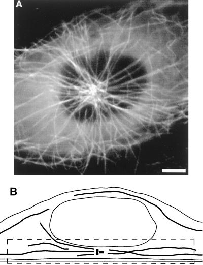
Figure 1.
Fluorescently labeled MTs in PtK1 cells. (A) Fluorescent MTs in a living PtK1 cell. The nucleus excludes MTs and tubulin monomers, creating a region in the center of the cell with little or no background fluorescence. In the cell shown here, the centrosome is in the center of this darkened region, making it easier to see single MTs. (Bar = 5 μm.) (B) Diagram depicting the geometrical relationship among the ventral cortex, the centrosome, MTs, and the nucleus as seen in side view; the components are not necessarily drawn to scale. The dashed lines show the approximate region of focus.

