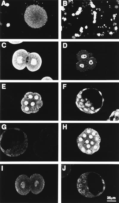
Figure 2.
Ontogeny of GH receptor immunoreactivity in preimplantation mouse embryos. Shown are confocal images of optical sections of the following. (A–F) Fertilized oocyte (A), cumulus cells (B), two-cell embryo (C), four-cell embryo (D), morula (E), and blastocyst (F) incubated with GH receptor antiserum. (G) Two-cell embryo and blastocyst incubated with preimmune serum. (H) Morula incubated with GH receptor antiserum preabsorbed with 20 μg/ml GST. (I and J) Two-cell embryo and blastocyst incubated with GH receptor antiserum preabsorbed with 20 μg/ml rabbit GH receptor. Note positive immunoreactivity appears on cumulus cells surrounding ovulated oocytes and is localized to the nuclei of cleavage-stage embryos and blastocysts. Consistent staining was observed in at least three experiments in which a total of 150 embryos were surveyed.

