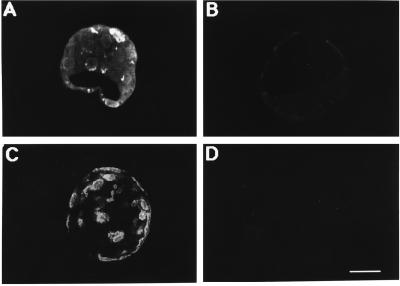
Figure 3.
Confocal images of mouse blastocysts showing positive immunoreactivity for GH. Blastocysts were incubated with monkey anti-rat GH antiserum (A and C), nonimmune monkey serum (D), or GH antiserum preabsorbed with 20 μg/ml rat GH (B). In a reconstructed three-dimensional image of a blastocyst (C) positive immunoreactivity appears on the outer membranes of the trophectoderm, with some immunoreactivity apparent in nuclei. In optical sections (A) the immunoreactivity is also apparent in cytoplasmic perinuclear vesicles. Consistent staining was observed in three experiments in which a total of 60 blastocysts were surveyed. (Bar = 25 μm.)

