
Figure 5. B cell activation in B6.ApoE-/- mice.
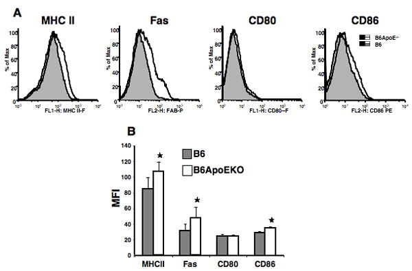
Splenic lymphocytes were analyzed at thirteen weeks post induction of cGVH. Cells were gated by size and CD19. (A) histograms from one representative mouse in each group; (B) means and SD. *, p<0.05, comparing B6.ApoEKO (N = 5 females) and B6 control (N = 3 females). MFI=Mean Fluorescence Intensity.