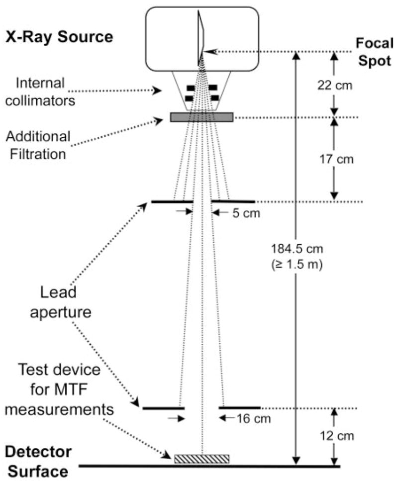
Figure 1.

DQE test geometry, compliant with the IEC 62220-1 standard. For the RQA5 beam quality, additional filtration with 21 mm of aluminum is used to simulate the spectral quality of radiation incident on the detector during a typical clinical examination. The detector is positioned at a source-to-image distance of 1.5 m or greater. The internal collimator of the device and external beam–limiting lead apertures are adjusted to achieve a radiation field of approximately 16 × 16 cm at the detector surface. The IEC standard specifies the exact position and size of only the aperture closest to the detector. The radio-opaque MTF device is placed adjacent to the detector as shown.
