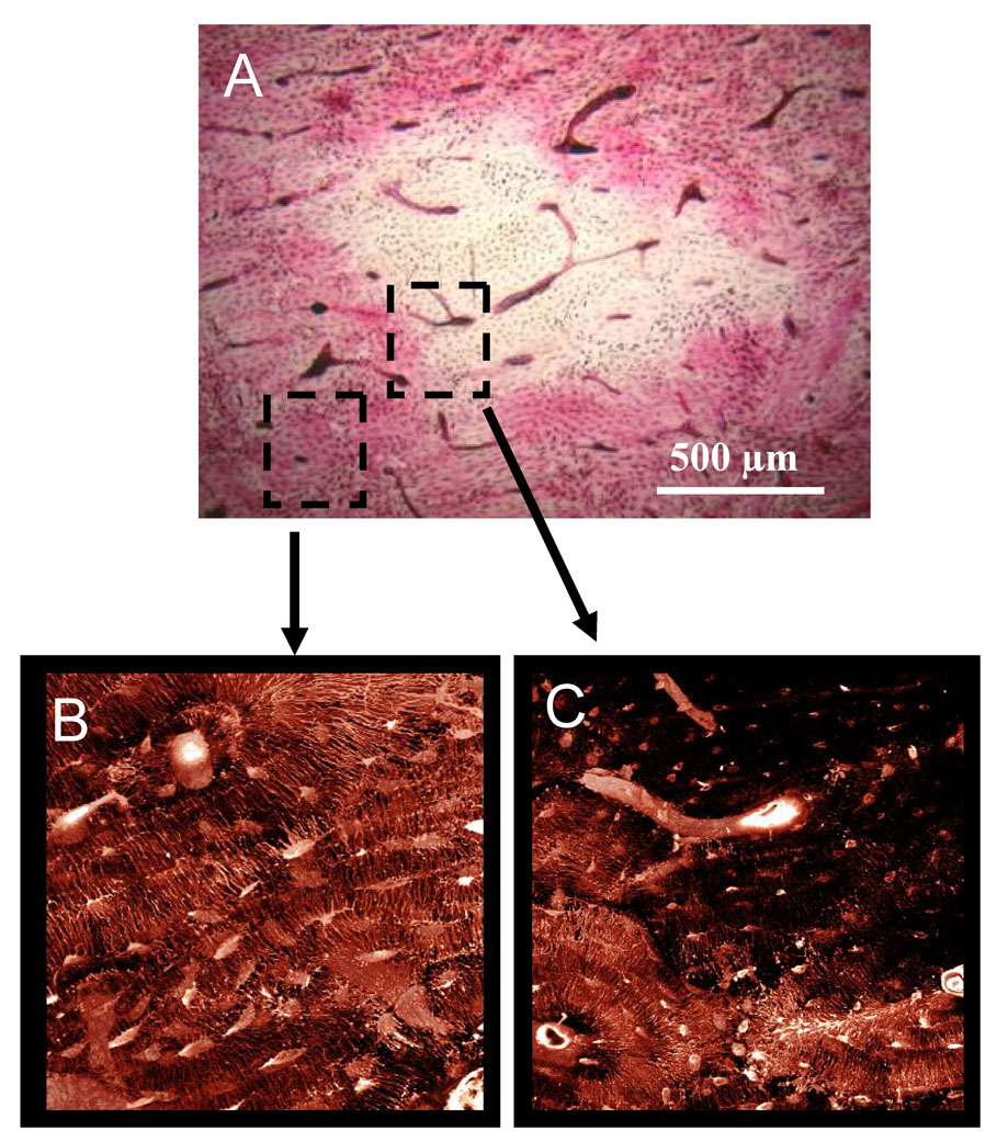
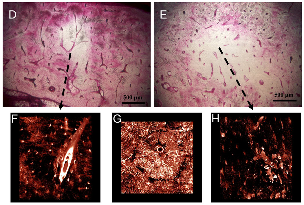
Figure 2.
Matrix necrosis in the mandible of dogs treated for 3 years with alendronate. The second molar region was stained en bloc with basic fuchsin with regions of matrix necrosis defined as those regions void of stain uptake. Necrotic bone matrix can be observed using brightfield microscopy (A, D, E). Due to its fluorescent properties, stained regions can be visualized using confocal microscopy, revealing patent canalicular networks (B,G). Conversely, necrotic regions which are void of stain can are without patent canalicular networks (C, F, H).


