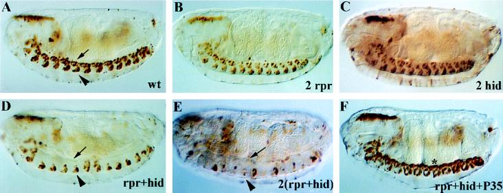
Figure 3.
rpr and hid act cooperatively to kill CNS midline cells. Shown are anti-β-galactosidase staining of stage 16 embryos bearing only P[52A-Gal4] and P[UAS-lacZ] (A), or additionally bearing two copies of P[UAS-rpr] (B), two copies of P[UAS-hid] (C), one copy of P[UAS-rpr] and one copy of P[UAS-hid] (D), two copies of P[UAS-rpr] and two copies of P[UAS-hid] (E), or one copy each of P[UAS-rpr], P[UAS-hid], and P[UAS-p35] (F). (A) P[52A-Gal4]/P[UAS-lacZ] embryos exhibited β-galactosidase expression in 2–3 midline glia (arrow) and the VUM neurons (arrowhead) in each segment. In embryos containing two copies of P[UAS-rpr] (B) or two copies of P[UAS-hid] (C), essentially normal numbers of midline glia and VUM neurons were present, indicating that these genes were not individually sufficient to kill midline cells. In embryos containing one copy each of P[UAS-rpr] and P[UAS-hid] (D), there was a dramatic loss of midline glia, although most of the VUM neurons remained present. In embryos containing two copies of P[UAS-rpr] and P[UAS-hid] (E), there was more severe loss of midline glia, as well as elimination of most of the VUM neurons. In embryos containing one copy of P[UAS-rpr] and P[UAS-hid] as well as one copy of P[UAS-p35], both ectopic and normal midline cell deaths were blocked. Note that many of the ectopic midline cells aggregated at the dorsal surface of the nerve cord (star). All views sagittal with anterior to left. (×100.)

