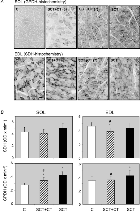
Figure 2. Histochemical changes in markers of energy metabolism in SOL and EDL muscles of the rat, induced by a complete spinal cord transection: effect of OEG transplantation.
Cross-sections of soleus and EDL muscles were subjected to quantitative glycerol-3-phosphate dehydrogenase (GPDH) or succinate dehydrogenase (SDH) histochemistry, as indicated in the Methods section. A, representative sections of sham-operated controls (C, n = 8) and spinal cord-transected (SCT, n = 11) rats, together with two examples of SCT rats transplanted with OEGs (SCT + CT, n = 9) showing a different degree of functional recovery (3 and 7, good and bad recovery, respectively (Ramon-Cueto et al. 2000)), stained for GPDH (soleus) or SDH (EDL). B, graphic representation of the quantitative histochemistry data for GPDH and SDH in both muscle types. Data are means ± s.d. of the number of animals per group indicated above, corresponding to 2-fold number of samples (both hind limbs) per group. *P < 0.05, SCT or SCT + CT versus sham-operated controls. #P < 0.05, OEG-transpanted (SCT + CT) versus non-transplanted SCT-rats. Bar (situated in the control EDL muscle for SDH histochemistry) indicates 1 μm.

