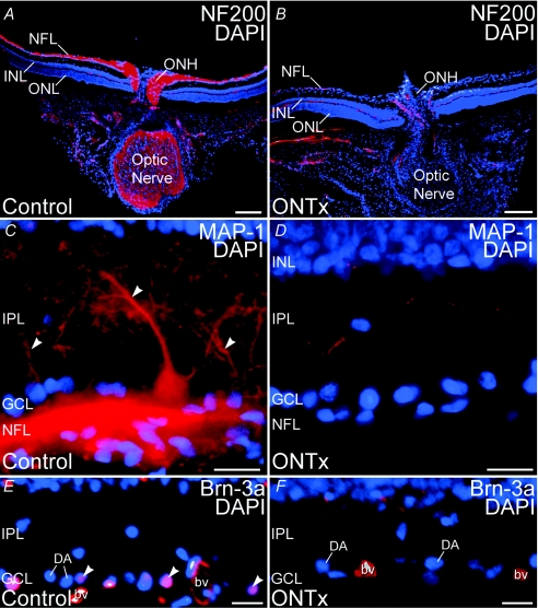
Figure 10. Optic nerve transection (ONTx) ablated retinal ganglion cells 3–6 weeks after surgery.
Immunolabelling for ganglion cell-specific markers is eliminated by ONTx. A and B, double labelling for neurofilament 200 kDa (NF200, red) and the nuclear dye DAPI (blue) in control retina (A) and retina 4 weeks after ONTx (B). C and D, double labelling for MAP-1 (red) and DAPI (blue) in control retina (C) and retina 6 weeks after ONTx (D). Large MAP-1-positive ganglion cell dendrites (arrowheads) are absent in the IPL after ONTx. E and F, double labelling for Brn-3a (red) and DAPI (blue) in the control retina (E) and retina 6 weeks after ONTx (F). Ganglion cell nuclei showing double labelling (arrowheads) are present in the control retina, but absent from ONTx retina. Displaced amacrine cells (DA) in the ganglion cell layer (GCL), however, persist in ONTx retina. Labelling in blood vessels (bv) is non-specific. ONL, outer nuclear layer; INL, inner nuclear layer; IPL, inner plexiform layer; NFL, nerve fibre layer. Scale bars: 200 μm for A and B; 20 μm for C–F.

