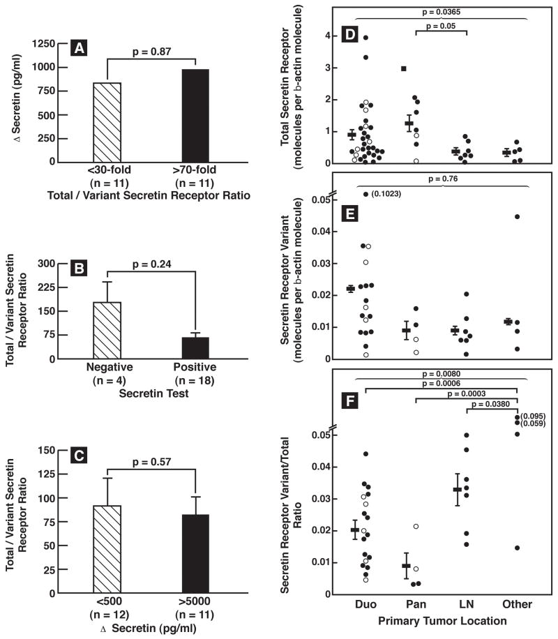
Figure 3.
Comparison of total-secretin and variant secretin-receptor expression or their ratio in gastrinomas with the positivity or magnitude of the secretin response or the tumor location. (Left Panel). The top panel compares the median Δsecretin in patients with gastrinomas that had <30-fold to those that had >70 fold greater amounts of total compared to secretin-receptor-variant abundance. The middle panel compares the total to variant secretin-receptor ratio in patients with or without a positive secretin test (i.e. >200 pg/ml increase). The lower panel compares the total to variant secretin-receptor ratio in gastrinomas from patients who had <500 pg/ml increase or >5000 pg/ml increase on secretin. (Right panel). Tumor location was determined at surgery. For the total-secretin-receptor expression results are from 31 duodenal, 7 pancreatic, 7 primary lymph node and 5 primary gastrinomas in other locations. For the variant and ratio results from 17 duodenal, 4 pancreatic, 7 lymph node primary and 4 primary gastrinomas in other locations are shown. Each dot represents the value from one gastrinoma in the indicated location and is a mean of at least 3 separate PCR determinations. Horizontal and vertical lines show the mean ± SEM for each primary tumor group.). The different symbols indicate the source of the tumor analyzed in each case. Symbols: ● Primary-sporadic, ○Primary-MEN1, ■LN-sporadic, □LN-MEN1, ▲Liver metastases
Abbreviations: Duod, duodenum; Panc, pancreas; Prim, primary; LN, lymph node.

