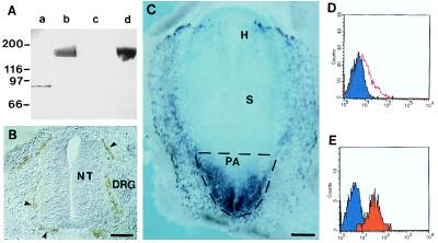
Figure 1.
Production of mAbs recognizing avian VEGFR2 and cell sorting of PA mesodermal cells. (A) Western blot with control CD4-Fc protein (10 μg, lanes a and c) and VEGFR2-Fc protein (10 μg, lanes b and d) was probed with anti-VEGFR2 (4H11) hybridoma supernatant (lanes c and d) and with anti-Fc mAb (lanes a and b). Anti-VEGFR2 recognizes only VEGFR2-Fc (lane d). (B) Immunohistochemistry with anti-VEGFR2 mAb on cryostat sections (20 μm) of an E4 chicken embryo. Section at the trunkal level showing anti-VEGFR2+ endothelial cells of the perineural vascular plexus (arrowheads). NT, neural tube; DRG, dorsal root ganglion. (Bar: 75 μm.) (C) Whole-mount in situ hybridization of a quail embryo at the 1-ss with a VEGFR2 antisense riboprobe. Note abundant positive cells in the mesoderm of the PA. H, headfold; S, somite (out of focus). The stippled region of the embryo was dissected. (Bar: 417 μm.) (D and E) Flow cytometry of VEGFR2+ cells from the PA. Fluorescence histogram showing anti-VEGFR2 labeling (red) before (D) and after (E) cell sorting.

