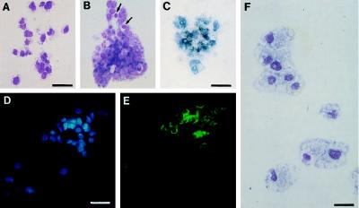
Figure 2.
Pheno types of hemopoietic cells obtained from VEGFR2+ PA cells. (A and B) MGG staining of hemopoietic colonies of thrombocyte (A) and thromboblast/erythroblast (B) type. Arrows in B point to two erythrocytes. (Bar: 25 μm.) (C) Benzidine-positive colony belonging to the erythrocytic lineage. (Bar: 21 μm.) (D) Hoechst nuclear stain and (E) CD 41/61 staining of a thromboblastic colony. (Bar: 26 μm.) (F) MGG staining of macrophages obtained in the presence of fibroblast-conditioned medium. (Bar: 18 μm.)

