
Figure 4. Spict regulates microtubules by inhibiting BMP signaling.
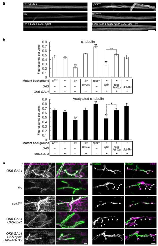
(a) Acetylated α-tubulin staining in segmental nerves passing through segment A4. (b) Quantification of levels of α-tubulin or acetylated-α-tubulin in 200-μm lengths of segmental nerve. Fluorescence intensity per voxel is normalized to a control channel (anti-HRP). Levels of anti-HRP are not significantly different among the genotypes in each panel (P > 0.05, n=6 larvae, one-way ANOVA). All comparisons are with w1118 unless indicated.. (c,i) Acetylated α-tubulin at NMJs of third instar larvae. Note that spictmut and tkv mutations affect both neuronal microtubules (overlapping with anti-HRP, green) and the muscle microtubule network visualized using anti-acetylated-α-tubulin (outside the anti-HRP-labeled area). Transgenes driven by the motor neuron-expressing OK6-GAL4 affect only presynaptic microtubules. (c,ii) Futsch is lost from distal boutons in tkv and in UAS-spict OK6-GAL4 NMJs (arrows show boutons labeled only with anti-HRP but not with anti-Futsch). Quantification is in Supplementary Fig. 4. In all panels, tkv mutant genotype is tkv7/tkv16713, tkvrescue is tkv7,OK6-GAL4/tkv16713; UAS-Tkv-HA. All images are projections of confocal sections. Scale bars, 10 μm.