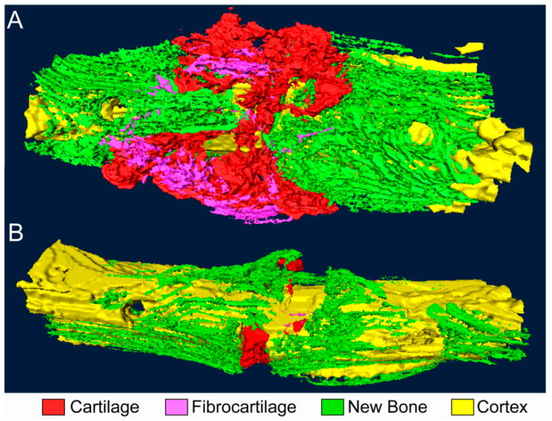
Fig. 3.

Bending stimulation of a full-thickness, transverse osteotomy gap. Three-dimensional reconstructions of serial histological sections of (A) a specimen following two weeks of bending stimulation and (B) a time-matched control that underwent continuous fixation with a four-pin external fixator. The mechanical stimulation induces formation of large amounts of cartilage within the gap and flaring outward toward the callus periphery in wedge-shaped configurations. Small amounts of fibrocartilage are found at the callus periphery, and bone formation is restricted to regions along the periosteal surface. No osseous bridging is observed. The control specimen exhibits bone formation within and surrounding the gap as well as a small amount of cartilage within the gap.
