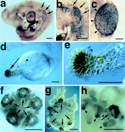
Figure 2.
Dll expression in representative deuterostomes. (a) Nine-day mouse embryo stained with the Dll antibody. Arrows (➞) point to medial border of cells expressing one or more Dlx genes in the presumptive forelimb. Dlx expression can be detected in developing mouse limbs as the bud forms from the flank, and somewhat earlier than previously reported for mice or other vertebrates (6–11). (b) Higher magnification view of the forelimb indicated in A. (c) Dorsal view of the forelimb of a 10-day mouse embryo stained with the Dll antibody. (➤) The position of the apical ectodermal ridge. (d) Three-day Molgula occidentalis ascidian larva from which an ampulla is extending. Cells at the distal tip of the ampulla express Dll (➞). (e) Higher magnification view of the ampulla shown in d. (f and g) Metamorphosing Strongylocentrotus droebachiensis sea urchin larvae stained with Dll antibody. Cells at the distal tip of the tube feet (➞) express Dll prior to (f) and during (g) extension from the body wall. (h) Higher magnification view of a tube foot (➞) and spines (➤) from an S. droebachiensis larva similar to that shown in g. Cells at the distal tip of the developing spines, as well as the tube feet express Dll. (Bars = 0.1 mm.)

