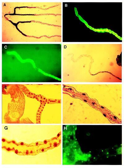
Figure 5.
Functional mapping of tubule regions. (A) Alkaline phosphatase staining of lower tubule. Tubules were fixed and stained for alkaline phosphatase. This shows a perfect overlap with line c507 (cf. Fig. 2G). (B and C) Short-term (10 min) labeling with 10 μg/ml rhodamine 123 identifies the main segment of both posterior (B) and anterior (C) tubules as the region responsible for transport of this organic molecule. (D) Initial and transitional segments express V-ATPase at a far lower level than the main segment, as detected by a reporter gene inserted into the vha55 gene in the P-element lethal line vha55j2e5 (28). This corresponds to the boundary reported by lines 155Y, c709, and c776 in Fig. 2 A and B. (E) A boundary midway along the ureter is marked by a change in the size of nuclei labeled in line vha55j2e5. This corresponds to the boundary reported by lines c649 and c601 in Fig. 2 H and I. (F) Within the main segment, only principal cell nuclei (cf. Fig. 4B) are labeled in line vha55j2e5. (G) Within the main segment, only principal cell nuclei (cf. Fig. 4B) are labeled in P-element line l(2)k02508, a recessive lethal insertion within the gene encoding the body-enriched vha68–2 V-ATPase A subunit (36). (H) The tiny cells (cf. Figs. 3F and 4C) are labeled by antibodies to horseradish peroxidase, which recognize an epitope on a Drosophila nervous system-specific Na+,K+ ATPase β-subunit (37).

