Figure 1.
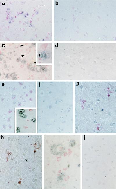
Expression of markers of oxidant stress (H0–1, a and b; p50, c and d), M-CSF (e and f), and VCAM-1 (i and j) in neurons proximate to Aβ, and expression of c-fms (g) in microglial in AD brain. a and c demonstrate increased expression of H0–1 and p50, respectively, in neurons of temporal lobe from AD brain, and b and d show weak staining for the same antigens in age-matched controls. The arrows and inset in c show nuclear localization of p50 in certain neurons. e and i display increased neuronal staining for M-CSF and VCAM-1 in AD brain, respectively, versus low levels of these antigens in age-matched controls (f and j, respectively). The inset in e shows location of M-CSF and plaques in double-stained AD brain (red is M-CSF; black is Aβ). g and h shows double immunostaining of the same section and depicts increased expression of c-fms in microglia in AD brain (g), identified by staining for CD68 (h); there is no staining for this antigen in age-matched controls (data not shown). These results are representative of immunohistologic analyses of five AD and four age-matched control brains (postmortem time of 2–6 hr). [Bar = 56 μm (a–f, j) and 12 nm (g–i).]

