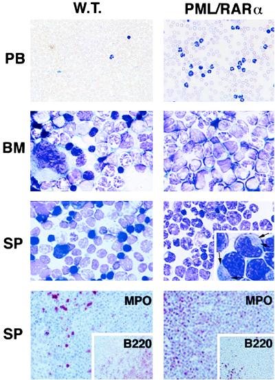
Figure 2.
Morphology and immunohistochemistry of PB, BM, and spleen (SP) cells from wild-type (W.T.) and leukemic hCG–PML/RARα transgenic mice. The smears of PB or imprints of BM and spleen were stained with Wright-Giemsa stain. Magnified detail of promyelocyte morphology is shown in the spleen imprint from the leukemic hCG–PML/RARα mouse. Auer’s bodies are visible in the cytoplasm of the leukemic promyelocytes as indicated by the arrows. The serial spleen sections shown in the bottom panel were immunohistochemically stained with MPO (myeloid precursors and mature granulocytes) and B220 (pan-B cells, Inset) antibodies. (×400 for PB; ×1,000 for BM and spleen; ×128 for immunohistochemical staining.) Note that in the leukemic mice, the number of WBC in PB is much increased; the normal BM components, including erythrocytes and megakaryocytes, are replaced by myeloid cells at various maturation stages; the spleen is heavily infiltrated by MPO-positive cells.

