Figure 4.
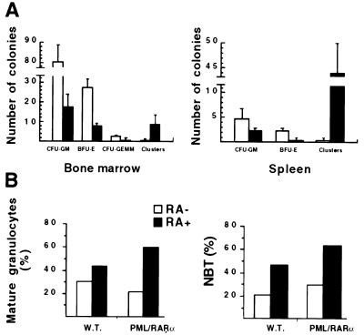
(A) Colony formation assay on BM and spleen cells from hCG–PML/RARα leukemic mice (solid bars) and sex- and age-matched wild-type littermates (open bars). Cells (3 × 10⁴) were plated per dish in 0.9% methylcellulose medium in triplicate. Colony numbers shown here are from one representative experiment (mean ± SD from the triplicate). CFU-GM includes CFU-G, -M, and -GM. Clusters are clones with <20 cells, often observed in assays performed on cells from acute leukemia and thought to represent colonies of leukemic origin (37). Note that colony formation from all lineages decreased and the number of clusters often observed in cultures from acute leukemias dramatically increased in BM and spleen cultures from leukemic transgenic mice. (B) In vitro response of BM cells to RA. BM cells from transgenic (PML/RARα) and wild-type (W.T.) mice were incubated with or without RA. Percentages of mature granulocytes and NBT-positive cells on day 5 are shown from two independent experiments. Note that both the percentage of mature granulocytes and the percentage of NBT-positive cells from leukemic transgenic mice are increased by 2-fold in the presence of RA.

