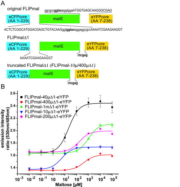
Figure 4.
Construction and in vitro analysis of improved maltose sensors. (A) Sketches of the original and improved versions of the FLIPmal sensors with shortened linkers. The amino acid sequence in capital letters corresponds to enhanced cyan fluorescent protein/enhanced yellow fluorescent protein or malE; small letters correspond to the synthetic linkers (sequence in bold is the restriction site). (B) In vitro fluorescence resonance energy transfer ratio changes of the FLIPmal sensor variants in the presence of maltose. Error bars represent the standard deviation (n = 3).

