
Figure 7. Accumulation of SCMAS in the brain of CLN2-mutant mice.
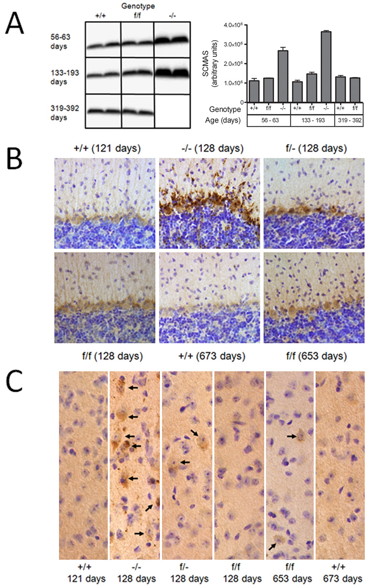
Panel A, Homogenates of mouse brain (10 ug protein equivalents) were fractionated by SDS-PAGE, transferred to nitrocellulose and SCMAS detected with a rabbit polyclonal antibody and visualized with an I125-labeled secondary antibody. Ages of congenic or near-congenic C57BL/6 mice were: +/+, 60, 63, 133 and 344 days; f/f, 59, 193, 319 and 392 days; −/−, 56 and 143 days. SCMAS levels in the mutant mice were determined from duplicate animals and error bars represent the range between these independent measurements. SCMAS was also detected by immunohistochemistry in the Purkinje cell layer of the cerebellum (Panel B) or cerebral cortex (Panel C). Arrows depicted individual neuronal cell bodies that stain for SCMAS. Age and genotype of animals are indicated for each panel.