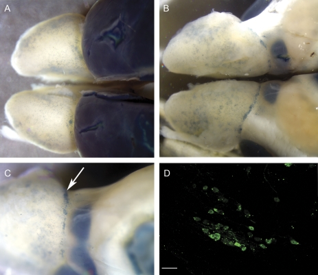
Figure 8.
Whole mount showing β-gal-reactive cells in blue. Notice the variability of β-gal-positive juxtaglomerular cell density throughout the OB surface. (A) Dorsal view of BDNFlacZneo mouse OBs and rostral cortex processed with X-gal reaction. (B) Ventral view. (C) Ventromedial view. Arrow indicates putative necklace glomeruli. (D) β-gal immunohistochemistry of putative necklace glomerulus. Scale bar: 20 μm.

