Figure 3.
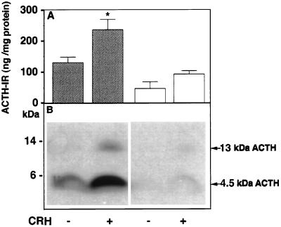
Radioimmunoassay and Western blot of immunoreactive ACTH in the secretion medium of anterior pituitary cells with and without CRH stimulation. (A) Secretion medium was subjected to HPLC and fractions were radioimmunoassayed using DP6 ACTH antiserum. Immunoreactive ACTH values corresponding to the retention times for ACTH1–39, including oxidized and basic-residue extended forms, were summed and expressed as ng ACTH-IR/mg cell protein, from which the medium was derived. The solid and open bars show the mean value ± SEM from three HPLC runs of medium from normal and Cpefat mouse pituitary cells, respectively, with and without CRH stimulation. ∗, P < 0.05 relative to control in normal mice. (B) Western blot analysis of secretion medium showing stimulated secretion of 13 kDa and 4.5 kDa ACTH with CRH in normal mice and low levels of 4.5 kDa that was not stimulated by CRH in Cpefat mice.

