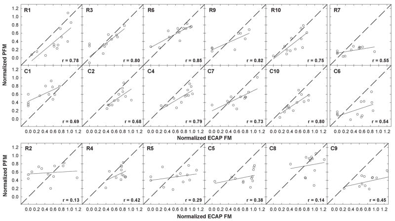
Fig. 2.

Individual-subject plots showing normalized psychophysical forward masking (PFM) plotted relative to normalized electrically evoked compound action potential forward masking (ECAP FM) for all masker-probe electrode pairs tested within a subject. Each panel represents data from a different subject; subject numbers are indicated in each panel. Normalization points (masker-equals-probe electrode) were removed and a correlation coefficient (indicated in each panel) was obtained. Solid lines represent results from linear regression analyses. Diagonal dashed lines represent unity. The top row shows data from Nucleus subjects with strong and moderate correlations. The middle row shows data from Clarion subjects with strong and moderate correlations (both subjects with moderate correlations are offset at right). The bottom row shows data from the remaining six subjects with poor correlations.
