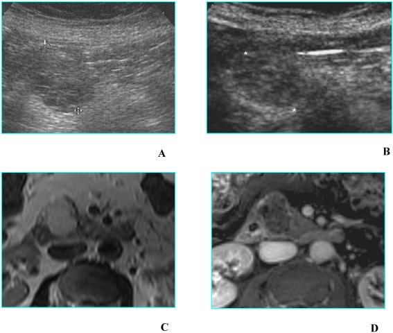
Fig 4.
Serous microcystic adenoma in an asymptomatic 66-year-old woman. A. B-mode US shows a hypoechoic mass (calipers) located in the head of the pancreas. B. On CEUS the lesion has a honeycomb appearance (asterisks), typical of serous microcystic adenoma. C. Axial T2-weighted MR image shows a hyperintese lesion made up of cluster of microcysts. D. Axial gadolinium-enhanced fat-suppressed T1-weighted MR image depicts the honeycomb appearance with enhancement of thin septa.

