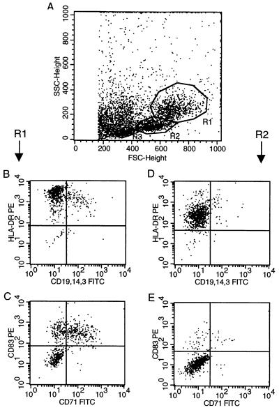
Figure 1.
Dot blot analysis of IMS-DCs isolated from peripheral blood. After a 24-h culture in the presence of granulocyte–macrophage colony-stimulating factor and IL-4, cell morphology (A) and protein expression (B–E) for mature DCs [region 1 (R1); B and C] and precursor DCs [region 2 (R2); D and E] were analyzed. For double immunofluorescence staining, mAb against HLA-DR and a mixture of CD3-, CD14-, and CD19-specific antibodies (B and D) or antibodies against CD83 and CD71 (C and E) were used.

