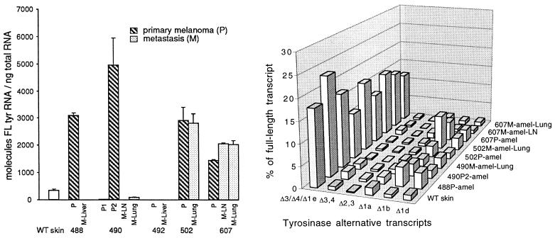
Figure 2.
Tyrosinase mRNA expression in amelanotic cutaneous melanomas and their amelanotic metastases, and in wild-type (WT) C57BL/6 skin. The numbered primary (P) melanomas, shown with their metastases (M) in lymph node (LN), lung, and liver, were analyzed as in Fig. 1. (Left) Absolute levels of the full-length tyrosinase mRNA relative to total RNA. Results are means and SD for two to five separate experiments. (Right) Levels of alternative mRNAs relative to the full-length mRNA in the same tumors. The most abundant alternative transcripts are represented by the bars, with the Δ3, Δ4, and Δ1e transcripts combined here. [The following tumors with minimal tyrosinase expression or none, as shown in the histogram (Left), are omitted in the diagram (Right): 488M-Liver, 490P1, 490M-LN, 492P, and 492M-Liver.]

