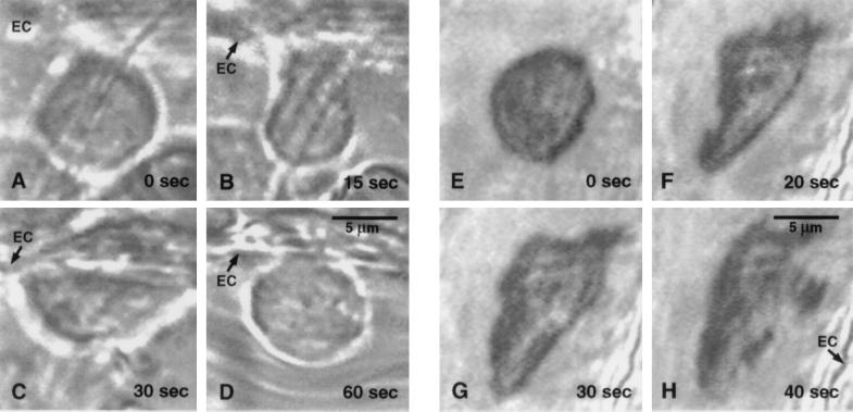
Figure 2.
Selected frames during the time course of pseudopod formation of a neutrophil in a rat mesentery venule. (A–D) Adhering neutrophil. (A) The leukocyte shape is spherical without pseudopods immediately after occlusion of the vessel with a micropipette; (B and C) leukocyte spreading on the endothelium (EC) by active pseudopod formation during stasis, (D) retraction of pseudopods upon restoration of flow. (E–H) Freely suspended neutrophil. (E) The cell is initially spherical. (F) Pseudopod projection shortly after flow stoppage, and continuation throughout stasis (G and H). The cell is carried away from the observation field upon return of flow.

